Stem cross-section
The plate shows the organisation of a fleshy stem. Green around the perimeter marks the photosynthetic region, and the pale tissue deeper inside marks the water storage region. The vascular system surrounds the pith. [Mauseth 2006]
This is a structural model. Tissue proportions, continuity of the hypodermal layer and distribution of additional bundles in the cortex vary among cacti.
Enlarge plate ↗Succulence and the division of functions in the shoot
The green surface of a cactus is part of the stem, which in many species has taken over much of the photosynthesis performed by leaves in other plants. The shoot interior contains extensive living tissues capable of storing water. This organization allows the plant's current functioning to continue through brief interruptions in rainfall. It does not imply an unlimited reserve. During drought, tissue hydration, volume and physiological activity gradually change. [Mauseth 2006]
A shoot cross-section reveals the epidermis, often a strengthening hypodermal zone, an extensive cortex, and a deeper vascular system and pith. Here, cortex is the name of a tissue, not the woody covering familiar from tree trunks. Its outer portion contains chloroplasts, while the more internal regions mainly participate in water storage. The boundaries and proportions of these zones depend on species, age and shoot type. [Mauseth 2006]
The cactus family also includes plants with well-developed leaves and epiphytes with relatively thin, flattened stems. A single cross-section of a globose cactus therefore cannot represent all its diversity. It is a model of one form of organization, useful for comparing plants, rather than a universal pattern for every member of Cactaceae. [Mauseth 2006]
Names of cactus parts
| Element | Meaning | How to interpret it |
|---|---|---|
| Shoot | The stem together with leaves and buds, even when leaves are highly reduced | The green body does not include roots. Shoot and stem are sometimes used as shorthand, but are not fully equivalent. |
| Growing tip | The region containing the shoot apical meristem | New sections of the axis and organ primordia form here. Not every areole is a growing tip. |
| Areole | A specialized short shoot with its own meristematic region | It can produce spines, hairs, lateral shoots or flowers. It is not a single spine or a hole in the epidermis. |
| Rib | A longitudinal ridge on the stem | Its geometry facilitates volume changes in the hydrated shoot. Rib number may change during growth. |
| Tubercle | A distinct projection on the shoot surface | It may be associated with an areole. Its arrangement and grooves matter in genus descriptions. |
| Axil | In mammillaria descriptions, the depression between tubercles | The flowering region may be separate from the spine-bearing apical portion of the areole. |
| Radial spine | A spine arranged radially near the areole's perimeter | It usually lies closer to the shoot surface. The distinction from a central spine concerns position, not just length. |
| Central spine | A spine developing nearer the centre of the spine cluster | It often projects farther from the surface. It may be straight or hooked. |
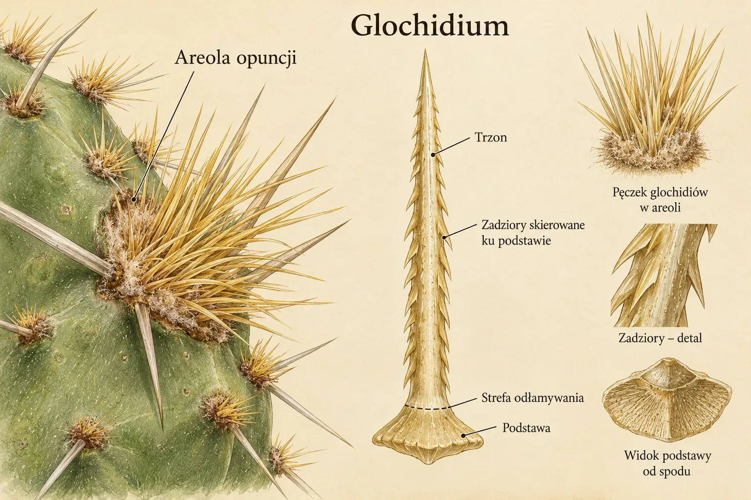
| Glochid | A small barbed spine typical of opuntioids | It detaches easily and penetrates skin. Glochids are not soft hairs that can be touched safely. |
| Areole hairs | Trichomes, often forming a woolly covering | They should be distinguished from thin or feathery spines. Similar appearance does not demonstrate shared structure. |
| Segment | A shoot section separated by a distinct constriction | A flat opuntia or schlumbergera segment is part of the stem, not a leaf. |
| Epidermis and cuticle | The covering tissue and its outer protective layer | Stomata belong to the epidermis. The cuticle limits water loss but does not seal the plant completely. |
| Photosynthetic parenchyma | Living cells with chloroplasts | It performs photosynthesis. It usually lies in the outer portion of the shoot cortex. |
| Water-storage tissues | Living tissues maintaining a water reserve | They are not an empty cistern. Storage and photosynthetic functions may partly overlap. |
| Xylem and phloem | Conducting tissues | Xylem carries water and minerals, while phloem mainly carries organic compounds between sites of production and use. |
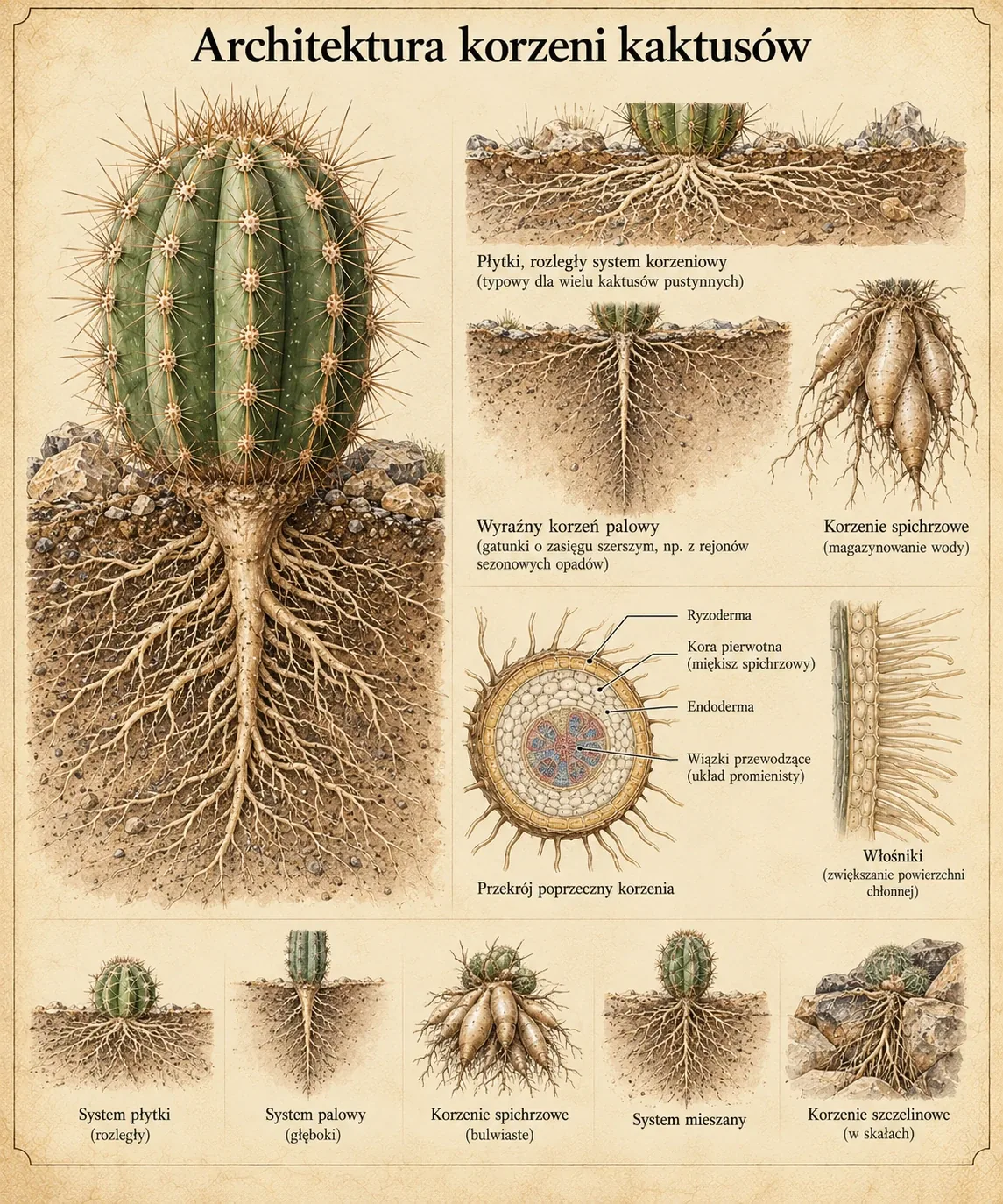
| Root collar | The transition zone between shoot and root system | Its position helps assess correct planting depth and the location of damage. |
| Primary root and lateral roots | The axis of embryonic origin and its branches | Roots arising from the shoot of a cutting are adventitious. A large root is not necessarily a primary root. |
| Storage root | A thickened root storing resources | It is not an underground stem. Cacti differ in the contribution of roots and shoots to water storage. |
Greek and Latin elements found in names are gathered in five nomenclature tables. The glossary describes language, while the comparison above helps identify organs and tissues. [Mauseth 2006] [Dubrovsky and North 2002]

Ribs, tubercles and stem form
Two rows of panels compare rib direction, number, width and degree of division into tubercles. These traits help describe stem appearance and changes during growth. [Mauseth 2006] [Hernández-Hernández et al. 2011]
The plate labels combine different descriptive criteria. Segmentation of the whole stem is not a rib type, and tubercles do not form a single simple evolutionary series from ribs. This comparison is not a formal classification.
Enlarge plate ↗The areole and growth organization
The areole is a specialized region associated with an axillary bud and a strongly shortened shoot. It usually groups spines and hairs, and its meristem may participate in forming further organs. A meristem is a tissue whose cells retain the ability to divide and give rise to new plant parts. Depending on species and developmental stage, lateral shoots or floral structures may arise from an areole. [Mauseth 2006]
The visible spine cluster thus reflects a specific developmental organization. The areole should not be equated solely with the place where spines are attached. In plants with reduced or invisible spines, areoles remain part of the shoot structure. Their presence helps distinguish a cactus from a similar-looking succulent belonging to another family. [Mauseth 2006]
In mammillarias, areole organization is particularly clear. The spine-producing portion lies at the tubercle tip, while activity associated with flowers or offsets occurs near its base. In collectors' descriptions, this region is called the axil between tubercles. Spatial separation of functions does not mean that the flower develops at a random location outside the plant's developmental system. [Mauseth 2006]

Structures developing in an areole
An areole is a strongly shortened lateral shoot. The plate compares a young areole, spine formation, flower bud development and an areole with mature spines. The panels help distinguish structures but do not define a mandatory order of appearance. In some cacti, a period of meristem dormancy separates spine formation from flowering. [Mauseth 2006]
The label “meristem” refers to the formative tissue, not the whole visible areole cushion. The 5 mm scale bar is not linked to an identified species or a measurement. The figure does not document an experiment or development time.
Enlarge plate ↗Origin of spines and hairs
Cactus spines are organs of leaf origin that develop on a short shoot. In developmental accounts, they are also compared with highly modified bud scales. Hairs covering the areole are epidermal outgrowths. Both structures may form a shared covering, but their similar appearance does not imply the same origin. [Mauseth 2006] [Silva et al. 2020]
A cactus spine and a rose prickle are also different organs. A prickle is a surface outgrowth, whereas a cactus spine is associated with the leaf developmental programme. Drawing a conducting strand inside one of them is not sufficient to explain the difference. In his 2006 review, Mauseth noted the absence of described vascular tissues and procambium in ordinary spines. However, the later study by Silva et al. 2020 demonstrated xylem and phloem at the base of specialized secretory glochids in two opuntioid species. Identifying a structure's origin rests on its development. The presence or absence of a vascular bundle is not a universal criterion for all spine modifications. [Mauseth 2006] [Silva et al. 2020]
A young spine has a region of division and elongation at its base. As it matures, most of its cells form a hard, lignified structure. Radial and central spines may differ in shape, thickness and growth direction. These features are useful for identifying plants, but need to be assessed alongside other properties and the age of the growth being examined. [Mauseth 2006] [Silva et al. 2020]

Areole and spine development
The left panel shows the positions of radial and central spines. The central panel connects the areole with stem tissues, and the right panel shows the elongated, hard cells of a mature spine. An areole is a short shoot that also produces hairs and other structures. [Mauseth 2006] [Silva et al. 2020]
The information about absence of xylem and phloem concerns ordinary spines. Specialised secretory glochids described in two species have a vascularised base and are shown on a separate plate.
Enlarge plate ↗Epidermis, cuticle and hypodermal zone
The epidermis separates the shoot interior from its surroundings. The cuticle and waxes covering it limit water loss but do not form a completely impermeable coating. Gas exchange occurs mainly through stomata. Their distribution is an anatomical feature, while their degree of opening changes in response to the plant's state and environmental conditions. [Mauseth 2006]
Many cacti have a hypodermis beneath the epidermis, a subepidermal zone of cells with thickened walls. It contributes to protection and the mechanical properties of the shoot surface. Its development varies, however. Some small cacti have a poorly developed hypodermis or do not form a distinct layer of this type. Cuticle thickness likewise provides no basis for simply ranking all species by drought resistance. [Mauseth 2006]
The green colour visible through the epidermis comes mainly from chloroplasts lying deeper within. Waxes, hairs and pigments also influence surface appearance. The waxy bloom is part of the plant's biological surface. It should be distinguished from deposits left by water or signs of pest infestation. [Mauseth 2006]
Photosynthetic and water-storage tissues
Photosynthetic parenchyma lies in the outer portion of the cortex. Light reaches it, and its chloroplast-containing cells perform photosynthesis. More internal tissues contribute more to water storage. Water is held in living cells, largely in their vacuoles. The shoot is not an empty tank surrounded by a green wall. [Mauseth 2006]
Different zones do not necessarily lose water at the same rate. Storage tissues can decrease in volume, helping maintain hydration in the photosynthetic layers. The relationship between cell structure and behaviour during drying is one reason why two similarly sized cacti may respond differently to prolonged water shortage. [Mauseth 2006]
The tissues of many species also contain mucilage cells. Mucilages are polysaccharide-rich substances that bind water strongly. Their presence affects the properties of hydrated tissues, but their contribution and the distribution of mucilage cells vary. Features of a single species should not all be extended to the entire family. [Mauseth 2006]
The vascular system in a thick stem
Xylem mainly conducts water and minerals, while phloem helps distribute photosynthetic products. In a thick stem, the distance between the green surface and the central vascular system itself becomes a significant problem. Long-distance transport cannot rely solely on the slow movement of substances between successive cells. [Mauseth 2006]
In many cacti, additional cortical bundles form a network within the extensive cortex. They bring conducting tissues closer to metabolically active regions and help supply a large volume of living shoot tissue. A diagram with a single strand running through the cactus centre is therefore insufficient. A simplified cross-section needs to distinguish the central bundle system from the additional cortical network. [Mauseth 2006]
The mechanical role of xylem also varies. A tall, columnar stem needs different support from a small globose plant. Not every cactus is equally soft inside, and a columnar habit involves more than elongating the structure characteristic of a small globose plant. [Mauseth 2006]
Ribs, tubercles and volume changes
The shoot expands after hydration and contracts during drought. Ribs partly compensate for these changes as surface folds spread apart or move closer together. The plant can thus change its cross-sectional shape without proportionally stretching the entire mature epidermis. Tubercles also participate in changes to shoot geometry, but their arrangement offers different possibilities for deformation from continuous ribs. [Mauseth 2006] [Hernández-Hernández et al. 2011]
Ribs affect both shoot illumination and mechanics. At any given moment, some surface areas receive strong sunlight while others are shaded. The result depends on rib height, shoot orientation and the covering formed by spines. Saying that ribs serve only shading or only water storage overlooks some of their functions. [Mauseth 2006] [Hernández-Hernández et al. 2011]
Earlier literature sometimes presented ribs and tubercles as successive steps along a single transformation pathway passing through modern genera. A comparison of shapes alone does not reconstruct evolutionary history. Modern species are the tips of a branching tree of relationships, not a series of ancestors and descendants set side by side. [Mauseth 2006] [Hernández-Hernández et al. 2011]
Interpreting plant structure in a collection
When observing a plant, distinguish persistent features from changes over time. Areole arrangement and tubercle type help describe shoot organization. The degree to which ribs spread apart may reflect current hydration. The form of the youngest growth also indicates the conditions in which it developed. An elongated, poorly coloured section should therefore be compared with older sections and the history of its growing position. [Mauseth 2006] [Fleischer and Schütz 1978] [1-WWW]
Shoot softness alone does not yield an unambiguous diagnosis. It may accompany the use of reserves, seasonal hydration changes or damage to tissues and roots. Combining anatomy with physiology, season and substrate conditions allows a meaningful interpretation of the observation. The following accounts of CAM and the root system develop this connection. [Mauseth 2006] [Fleischer and Schütz 1978] [1-WWW]
Areole tissues and regeneration after chilling
Meristems of areole axillary buds may retain viability at temperatures that more severely damage photosynthetic parenchyma. Zutta and co-authors compared tissues of three cultivated cacti after 100 days at 10/5°C. In Opuntia robusta, LT₅₀ was −8.8 ± 0.4°C for parenchyma and −11.2 ± 0.4°C for the meristem. Means ± standard error were given for five plants after one hour of exposure. Meristem samples came from areoles, which must be distinguished from examining the main growing tip. [Zutta et al. 2011]
Greater bud tolerance is one possible basis for regrowth after shoot-surface damage. Cell survival in a test is not sufficient to demonstrate later regeneration of the entire plant. Regeneration experiments also need to examine bud and root development. Data on meristems and regeneration. [Zutta et al. 2011] [Ishikawa and Gusta 1996]
Tubercle types and species examples
Tubercle shape is described independently of density and spine structure. The short cones of Mammillaria mammillaris, elongated tubercles of M. longimamma and lateral flattening in M. compressa illustrate distinct features. Eight plates with examples and commentary on terminology allow comparison without treating the diagrams as an identification key. [Britton and Rose 1923] [2-WWW] [2-WWW] [2-WWW]
Chlorenchyma of a persistent opuntioid leaf
In Austrocylindropuntia subulata, the cylindrical leaf contains an outer palisade region and an inner non-palisade region, both photosynthetically active and storing water. In the material studied, mucilage idioblasts were scattered between them. Leaf structure can be compared with measurements of its daily CO₂ exchange, distinguishing anatomical from physiological data. [Ozerova and Timonin 2020b]
Cholla wood and wound remodelling
Cylindropuntia imbricata forms openwork wood with several scales of porosity. The development of a barrier and wound periderm was studied in C. bigelovii and Opuntia ficus-indica. The account of cholla wood describes the skeletal structure, while successive stages of regeneration are presented in How a cactus closes a wound. [Morankar et al. 2024][Mylo et al. 2020]
New ribs near the apex
Ribs were counted on 100 randomly selected plants in a Ferocactus acanthodes population at Deep Canyon. In 56%, the number in the middle of the stem was 5, 8, 13 or 21. New ribs appeared nearer the apex, and intermediate values occurred mainly in this region. Rib number therefore changed in stages associated with growth. The authors interpreted this as a way of adjusting the geometry of a larger stem to volume changes during fluctuations in hydration. The mechanism triggering successive stages remained unknown. [Robberecht and Nobel 1983, pp. 153–155]
The anatomical explanation is based on the full text of Mauseth's 2006 review and the Structure and Forms sections of the old site. Historical terms are compared with the modern developmental description. The diagrams show relationships between tissues without preserving their relative thicknesses. The account of spine structures is supplemented by the vascularized base of secretory glochids according to Silva et al. 2020.
Budowa, fizjologia i rozmnażanie kaktusów
[Autor nieustalony, Budowa, fizjologia i rozmnażanie kaktusów, b.d.f]
Material from an older website, preserved in Konrad Szychowski's private archive. It covers cactus structure, physiology and propagation.
Structure–Function Relationships in Highly Modified Shoots of Cactaceae
Mauseth J. D., Structure–Function Relationships in Highly Modified Shoots of Cactaceae, Annals of Botany, 2006, 98(5):901-926, doi: 10.1093/aob/mcl133
The review describes cactus stem structure, areoles, and photosynthetic and storage tissues. It explains connections between anatomy, photosynthesis and water storage.
Kaktusy
[Fleischer Z., Schütz B., Kaktusy, Państwowe Wydawnictwo Rolnicze i Leśne, Warszawa, 1978, wyd. I, trans. Kabiesz Z., Prajer Z.]
Państwowe Wydawnictwo Rolnicze i Leśne, Warsaw, first edition. Translated by Zofia Kabiesz and Zbigniew Prajer. Historical and cultivation background. Names and interpretations updated separately.
Structure and function of secretory glochids and nectar composition in two Opuntioideae (Cactaceae) species
Silva S. C. D. M., Machado S. R., Nepi M., Rodrigues T. M., Structure and function of secretory glochids and nectar composition in two Opuntioideae (Cactaceae) species, Botany, 2020, 98(8):425-437, doi: 10.1139/cjb-2020-0004
The paper compares glochids of four Brasiliopuntia brasiliensis and five Opuntia cochenillifera plants (Nopalea cochenillifera in the publication). Microscopy, histochemistry and HPLC describe secretory function and xylem and phloem at the glochid bases.
Archiwalne zestawienia terminologii oraz schematy fizjologii, sukulentyzmu i rozmnażania
[Autor nieustalony, Archiwalne zestawienia terminologii oraz schematy fizjologii, sukulentyzmu i rozmnażania, b.d.a]
A set of tables and illustrations from Konrad Szychowski's private archive. It covers terminology, physiology, succulence and propagation. One figure refers to W. Nultsch's 1973 account, identifying that drawing's source rather than dating the entire collection.
Low- and High-Temperature Tolerance and Acclimation for Chlorenchyma versus Meristem of the Cultivated Cacti Nopalea cochenillifera, Opuntia robusta, and Selenicereus megalanthus
Zutta B. R., Nobel P. S., Aramians A. M., Sahaghian A., Low- and High-Temperature Tolerance and Acclimation for Chlorenchyma versus Meristem of the Cultivated Cacti Nopalea cochenillifera, Opuntia robusta, and Selenicereus megalanthus, Journal of Botany, 2011, 2011:347168, doi: 10.1155/2011/347168
The study compares the viability of photosynthetic parenchyma and axillary bud meristems in three cultivated cacti. It includes one hour of exposure to low temperature after different acclimation conditions.
Freezing and heat tolerance of Opuntia cacti native to the Canadian prairie provinces
Ishikawa M., Gusta L. V., Freezing and heat tolerance of Opuntia cacti native to the Canadian prairie provinces, Canadian Journal of Botany, 1996, 74(12):1890-1895, doi: 10.1139/b96-226
Seasonal studies of material from Saskatchewan examine cladode freezing, subsequent necrosis and regeneration. They compare hardened winter cladodes with young growth.
The Cactaceae: Descriptions and Illustrations of Plants of the Cactus Family. Volume IV
[Britton N. L., Rose J. N., The Cactaceae: Descriptions and Illustrations of Plants of the Cactus Family. Volume IV, Carnegie Institution of Washington, Washington, 1923, tom IV]
Volume IV of the 1923 monograph contains descriptions of cacti, including tubercle structure and historical name combinations. The supplied copy comes from Konrad Szychowski's private archive.
Plants of the World Online — Royal Botanic Gardens, Kew
Accepted names, synonyms and distribution. A citation beside a taxon leads to its record.
Accessed 2026-10-08.
Structure of the chlorenchyma in the persistent terete leaves of Austrocylindropuntia subulata (Muehl.) Backeb. (Cactaceae): anatomy beyond functional limitation
Ozerova L. V., Timonin A. C., Structure of the chlorenchyma in the persistent terete leaves of Austrocylindropuntia subulata (Muehl.) Backeb. (Cactaceae): anatomy beyond functional limitation, Wulfenia, 2020, 27:221-232
A microscopic analysis of the persistent, cylindrical leaves of Austrocylindropuntia subulata. It describes outer palisade parenchyma and inner non-palisade parenchyma, mucilage idioblasts and the arrangement of vascular bundles. The discussion concerns the combination of assimilation and water storage in the same tissues.
Cholla cactus wood (Cylindropuntia imbricata): Hierarchical structure and micromechanical properties
Morankar S., Luktuke A., Nieto-Valeiras E., Mistry Y., Bhate D., Penick C. A., Chawla N., Cholla cactus wood (Cylindropuntia imbricata): Hierarchical structure and micromechanical properties, Acta Biomaterialia, 2024, 174:269-280, doi: 10.1016/j.actbio.2023.12.005
Tomography, microscopy, nanoindentation and modelling of dry Cylindropuntia imbricata wood. The study describes structure from the openings in the skeleton to cell walls, and compares the properties of fibres and wood rays.
Self‐repair in cacti branches: Comparative analyses of their morphology, anatomy, and biomechanics
Mylo M. D., Krüger F., Speck T., Speck O., Self‐repair in cacti branches: Comparative analyses of their morphology, anatomy, and biomechanics, International Journal of Molecular Sciences, 2020, 21(13):4630, doi: 10.3390/ijms21134630
A comparison of the healing of controlled wounds in Cylindropuntia bigelovii and Opuntia ficus-indica over 31 days. Microscopy and bending tests show successive stages of protective barrier and wound periderm formation, and changes in stem stiffness.
A Fibonacci Sequence in Rib Number for a Barrel Cactus
Robberecht R., Nobel P. S., A Fibonacci Sequence in Rib Number for a Barrel Cactus, Annals of Botany, 1983, 51(1):153-155, doi: 10.1093/oxfordjournals.aob.a086440
The paper describes rib number and addition in Ferocactus acanthodes in Deep Canyon, California. In a sample of 100 randomly selected plants, rib number was compared with stem height and the distribution of ribs between the base and apex. The authors identified frequent Fibonacci sequence values and transitional phases. The link between this pattern, stem development and hydration changes was presented as a functional interpretation.
Root Structure and Function
[Dubrovsky J. G., North G. B., Root Structure and Function, Cacti: Biology and Uses, University of California Press, Berkeley i Los Angeles, Nobel P. S. (red.), 2002, s. 41–56]
The chapter presents cactus root structure and function. It discusses anatomy, water uptake and hydraulic conductivity under changing hydration conditions.
Phylogenetic relationships and evolution of growth form in Cactaceae
Hernández-Hernández T., Hernández H. M., De-Nova J. A., Puente R., Eguiarte L. E., Magallón S., Phylogenetic relationships and evolution of growth form in Cactaceae, American Journal of Botany, 2011, 98(1):44-61, doi: 10.3732/ajb.1000129
The molecular analysis presents cactus relationships and growth form evolution. Reconstruction indicates repeated origins of similar forms in different lineages.
Royal Horticultural Society
Cultivation and plant protection guides. Horticultural recommendations are read with attention to species and cultivation conditions.
Accessed 2026-10-08.