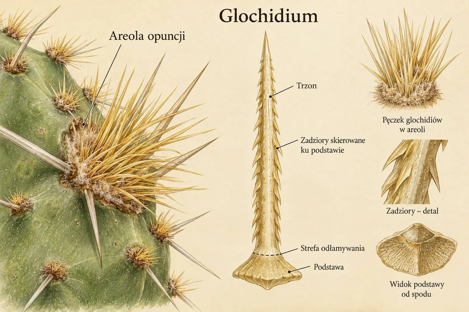
Opuntia glochidium
The large panel at left indicates the position of glochids in an areole. The central panel shows a single glochid with barbs pointing towards its base. At right are enlarged views of a bundle, the shaft surface and the base. [Robinson 1974]
The drawing is a schematic enlargement without a measurement scale. Base shape and the arrangement of surface cells are not identical in all opuntioids.
Enlarge plate ↗Spine development in the areole
A cactus spine is a structure of leaf origin developing in the areole, a specialized system associated with an axillary bud. Areole hairs arise from the epidermis. A shared location and similar protective function do not make them the same organ. A spine is also not equivalent to a rose prickle, an outgrowth of surface tissues. [Mauseth 2006]
In a developing spine, cellular activity at the base enables elongation. As it matures, many cell walls thicken and lignify. A hard mechanical structure forms. Lignification means lignin deposition in the cell wall, not merely drying of a living organ. A mature spine can therefore retain its shape long after growth ends. [Mauseth 2006]
Spine number, arrangement and shape can be important identification features. Growth of similar age should be compared. A young, developing spine may differ in colour and stiffness from an older one. Central and radial spines are distinguished by position in the areole, not as two entirely independent organ types. [Mauseth 2006]
The glochid and its detachment point
Glochids are small spine structures characteristic of opuntioids. They often form clusters that resemble soft fuzz from a distance. Their surface may nevertheless bear numerous barbs pointing towards the base. The sharp tip facilitates entry into tissue encountered, while the backward projections increase resistance to withdrawal. [Robinson 1974]
The base is equally important. In his 1974 paper, Robinson noted that easy detachment is associated with a zone that does not attain the same degree of sclerification as the rest of the structure. Sclerification is tissue hardening through wall thickening, usually accompanied by lignification. The difference between a glochid and a firmly attached spine therefore includes its attachment, not just length. [Robinson 1974]
Barbs are shaped portions of surface cells. They should not be imagined as separate hooks glued to a smooth needle. Their outlines are enlarged in the diagram to show their direction of action. Actual projection density and basal structure depend on the material examined. [Robinson 1974]
Glochid structure in Robinson's studies
Robinson compared spines and glochids from different opuntioids using a scanning electron microscope. Some material came from herbarium collections. He supplemented observations with light microscopy and inspection of bases under a stereomicroscope. Combining several methods distinguished actual shape from what is visible only with one orientation of a preparation. [Robinson 1974]
Among the bases, he identified flattened forms with lateral portions resembling a fish tail, and more rounded, expanded forms. Depressions or a channel extending into the interior were visible in some material. These are much subtler features than the general presence of barbs. They show that mechanically similar structures may differ in organizational details. [Robinson 1974]
The author also used these observations to discuss generic boundaries. Current classification should not be copied from the system adopted in 1974. The value of microscopic documentation and the currency of taxonomic conclusions are separate matters. On this site, the older paper is used to explain structure, while names and relationships are checked separately. [Robinson 1974]
Shoot protection and diversity of functions
Spines can restrict herbivore access to hydrated tissues. Their arrangement also changes the radiation reaching the shoot surface. A dense covering partly shades green tissue but also reduces the light available for photosynthesis. The significance of this feature therefore depends on habitat conditions, covering density and plant physiology. [Mauseth 2006] [Robinson 1974]
Spine colour, length and density do not form a simple ranking of drought resistance. Similar appearance may result from different combinations of traits. A rigid covering arranged around the shoot functions differently from a layer of hairs or a cluster of small glochids. Inference about function should combine structural description with measurement or observation of its action. [Mauseth 2006] [Robinson 1974]

Spines. Structure and possible functions
Panels 1–3 organise spine structure and descriptive traits. Panel 4 compares possible protective functions and effects on the environment around the stem surface. Panel 5 illustrates the developmental relationship between spines and leaves. [Mauseth 2006] [Robinson 1974] [Ju et al. 2012]
The arrow from leaf to spine indicates homology, not transformation of a mature leaf on a particular plant. The 100 μm scale bar is part of the drawing, with no measurement material specified. Spine effects on water and microclimate depend on species and conditions.
Enlarge plate ↗Droplet transport on Opuntia microdasys
In 2012, Ju and co-authors studied microstructures of Opuntia microdasys. They used the term spines in their paper. For this species' small, barbed structures, their relationship with glochids is important. The researchers observed a conical shape, longitudinal grooves and hairs at the base. They combined electron microscopy with filming droplet behaviour. [Ju et al. 2012]
The experiment used a fog stream generated by an ultrasonic humidifier with purified water. Flow speed was about 20–30 cm per second. Individual structures were placed at different angles. Droplets moved towards the base even when this required movement against gravity. Flow under their own weight alone could not explain the observations. [Ju et al. 2012]
Successive small droplets settled on the surface and merged into larger ones. Near the base, they contacted hairs between which capillary forces acted. Rapid removal of a droplet from the surface allowed another water-collection cycle to begin. This is a sequence of deposition, merging, movement and droplet capture, rather than a single process generally called absorption. [Ju et al. 2012]
Geometry and surface wettability
A cone has different radii near its tip and base. A droplet surrounding such a section rests on a surface of varying curvature. The authors identified a Laplace-pressure difference as one force favouring movement towards the broader part. A change in wetting properties associated with groove distribution was the second component of their explanation. [Ju et al. 2012]
The longitudinal arrangement of surface irregularities can make movement along the axis easier than across it. Asymmetric barbs further differentiate movement directions. In this interpretation, the shape of the whole structure, surface relief and water contact with hairs work together. None of these details is a biological pump by itself. [Ju et al. 2012]
Fog contains suspended droplets of liquid water. This experimental result does not mean spines absorb arbitrary amounts of water vapour from dry air. Directional transport was shown in a specific laboratory system. Assessing fog's contribution to the whole plant's water balance would also require data on field conditions, the amount of water collected and its subsequent route. [Ju et al. 2012]
Secretory glochids in two species
Silva, Machado, Nepi and Rodrigues described another specialization. They examined four adult Brasiliopuntia brasiliensis plants and five plants named Nopalea cochenillifera in the publication. Following POWO, the second species is written here as Opuntia cochenillifera. Material came from cultivation in Botucatu, Brazil. Young stem segments and successive developmental stages of areole structures were analysed. [Silva et al. 2020]
Hairs, ordinary spine structures and secretory glochids coexisted in a single areole. The latter were soft, translucent and larger than non-secretory glochids. Droplets appeared at their tips. An individual structure remained active for two or three consecutive days. This is a short developmental phase that can easily be missed during occasional collection inspections. [Silva et al. 2020]
The researchers used light microscopy, scanning and transmission electron microscopy, and histochemical reactions. Each method addressed a different question. Surface images revealed shape, sections showed tissue organization, and ultrastructure and staining helped interpret activity. [Silva et al. 2020]
The secretory base and nectar pathway
Active glochids had an expanded base, an elongated middle portion and a tapered tip. The base contained living parenchyma cells with abundant cytoplasm and xylem and phloem elements. Conducting tissues were confined to this portion. This is a significant exception to the simplified view of a spine as a dead, unvascularized structure that is uniform throughout its length. [Silva et al. 2020]
Based on cell organization, the authors assigned nectar production to the base, transport to the middle portion and release to the tip. Numerous plasmodesmata indicated possible movement through connected cytoplasms. Cell-wall structure was also consistent with an extracellular route. This interpretation rests on anatomy and cytochemistry, not a film showing every sugar molecule moving through the organ. [Silva et al. 2020]
No open pores or modified stomata were found on the surface. The authors inferred that secretion passed through the intact cuticle. After secretion ended, tissues in the middle and apical portions lignified more strongly. Function therefore depended on both species and the life stage of a particular glochid. [Silva et al. 2020]

Secretory glochids
The left side compares structures in Brasiliopuntia brasiliensis and Nopalea cochenillifera, accepted in the atlas as Opuntia cochenillifera. The right side distinguishes the expanded, vascularised base, unlignified middle part and tip. The drop at the tip indicates secreted nectar. [Silva et al. 2020]
The plate concerns specialised structures in two species studied. It does not show the structure of all glochids or ordinary spines. It is an educational reconstruction, not a microscopic image from a publication.
Enlarge plate ↗Secretion composition and ant presence
Chromatographic analysis detected glucose, fructose and sucrose. Sucrose accounted for about 88 percent of the sugars measured in B. brasiliensis and about 90 percent in O. cochenillifera. These percentages describe its share of the sugar pool, not its share of the entire droplet. Amino acids were also present in the secretion. Total sugar content was higher in the second species, while amino acid content was higher in the first. [Silva et al. 2020]
Ants collected secretion from glochid tips. This observation confirms that the structures provide food accessible to insects. Deterrence or attack of herbivores by visiting ants is a possible benefit to the plant. Insect presence alone does not measure the effectiveness of such protection. The authors identified a need to further test benefits to both partners. [Silva et al. 2020]
The result concerns two examined species and specific young structures. It does not demonstrate that every spine secretes nectar or that every glochid cluster has identical anatomy. A nectar droplet and a fog droplet may occur in the same shoot region, but arise and move through different processes. [Silva et al. 2020]
Documenting structures in a collection
Record the species, segment age, areole position and date when observing. A photograph repeated after several days may reveal a change absent from a single image. Comparing young and old areoles is particularly important when seeking brief secretory phases. [Chmielewski b.d.] [Robinson 1974]
Marian Chmielewski's archival text on spines and seeds rightly emphasizes the importance of a scale bar in a micrograph. A magnification label alone loses its meaning when the image size changes on screen. A scale bar shrinks or enlarges with the photograph, preserving information about dimensions. An educational diagram without a scale instead shows relationships and is not intended for measurements. [Chmielewski b.d.] [Robinson 1974]
Glochids detach easily during plant handling. A fluffy appearance is no basis for judging whether they are safe to touch. Optical observation is sufficient to document structure, without testing attachment on skin or removing structures from the plant. [Chmielewski b.d.] [Robinson 1974]
Directional movement of dew along a spine
In Copiapoa gigantea (C. cinerea var. haseltoniana in the study), droplet movement towards the spine base was recorded, including against gravity. The authors linked it to conical geometry, microgrooves and changes in wetting. The experimental account and mechanism diagram present the scope of the measurements. [Malik et al. 2016]
Spine covering, light and stem detachment
In Cylindropuntia bigelovii, the spine covering changes the light reaching the green stem, while easily detached segments participate in reproduction. The study of light physiology compares night-time acid accumulation, while mechanical experiments explain the role of geometry and tissue properties in segment separation. [Nobel 1983][Mylo et al. 2022]
Droplets on opuntia glochids
In Opuntia microdasys, a laboratory fog stream revealed successive deposition of small droplets, merging and movement towards the glochid base. Conical geometry, longitudinal grooves, barbs and differences in wettability work together, while dense areole hairs capture incoming water. Transport towards the base was also observed with an orientation opposing gravity. [Ju et al. 2012]
A similar system was described in O. stricta. The 2020 experiment used mist from a humidifier despite the title's reference to dew. Both papers establish the mechanics of liquid water on glochid surfaces. Quantifying this pathway's contribution to the field water balance requires separate measurements. [Masrahi 2020][Ju et al. 2012]
Glochids on young and older segments
Areole appearance changes with segment age. Griffiths described new glochids appearing in the central portion of the areole and their concentric arrangement in Opuntia xanthoglochia. In O. lubrica, he observed areolar tissue growing into short elevations densely covered with successive bundles. These morphological observations from the original descriptions document accumulation of structures on the same areole in successive seasons. [Griffiths 1910, pp. 166, 169]
The basis is the full texts of Ju et al. and Silva et al., together with the read scan of Robinson's paper, pages 278–283. Marian Chmielewski's archival text supplements microscopic documentation. The names Opuntia microdasys, Brasiliopuntia brasiliensis and Opuntia cochenillifera were checked in POWO. The diagrams are independent representations of biological relationships.
Structure–Function Relationships in Highly Modified Shoots of Cactaceae
Mauseth J. D., Structure–Function Relationships in Highly Modified Shoots of Cactaceae, Annals of Botany, 2006, 98(5):901-926, doi: 10.1093/aob/mcl133
The review describes cactus stem structure, areoles, and photosynthetic and storage tissues. It explains connections between anatomy, photosynthesis and water storage.
Scanning electron microscope studies of the spines and glochids of the Opuntioideae (Cactaceae)
Robinson H., Scanning electron microscope studies of the spines and glochids of the Opuntioideae (Cactaceae), American Journal of Botany, 1974, 61(3):278-283, doi: 10.1002/j.1537-2197.1974.tb10775.x
The paper presents glochid microscopic structure. It compares shaft surfaces, barbs and bases and discusses their significance in the systematics of the time.
A multi-structural and multi-functional integrated fog collection system in cactus
Ju J., Bai H., Zheng Y., Zhao T., Fang R., Jiang L., A multi-structural and multi-functional integrated fog collection system in cactus, Nature Communications, 2012, 3(1):1247, doi: 10.1038/ncomms2253
The study analyses water collection and droplet transport on Opuntia microdasys surfaces. It links observed movement to the geometry and microstructure of plant elements.
Structure and function of secretory glochids and nectar composition in two Opuntioideae (Cactaceae) species
Silva S. C. D. M., Machado S. R., Nepi M., Rodrigues T. M., Structure and function of secretory glochids and nectar composition in two Opuntioideae (Cactaceae) species, Botany, 2020, 98(8):425-437, doi: 10.1139/cjb-2020-0004
The paper compares glochids of four Brasiliopuntia brasiliensis and five Opuntia cochenillifera plants (Nopalea cochenillifera in the publication). Microscopy, histochemistry and HPLC describe secretory function and xylem and phloem at the glochid bases.
Plants of the World Online — Royal Botanic Gardens, Kew
Accepted names, synonyms and distribution. A citation beside a taxon leads to its record.
Accessed 2026-10-08.
Ciernie i nasiona kaktusów
[Chmielewski M., Ciernie i nasiona kaktusów, b.d.]
Marian Chmielewski's text preserved in Konrad Szychowski's private archive. It discusses spines and seeds and their microscopic documentation with a scale. Publication date remains undetermined.
Hierarchical structures of cactus spines that aid in the directional movement of dew droplets
Malik F. T., Clement R. M., Gethin D. T., Kiernan M., Goral T., Griffiths P., Beynon D., Parker A. R., Hierarchical structures of cactus spines that aid in the directional movement of dew droplets, Philosophical Transactions of the Royal Society A, 2016, 374(2073):20160110, doi: 10.1098/rsta.2016.0110
The authors track droplet movement along the spines of Copiapoa gigantea, described in the paper as C. cinerea var. haseltoniana. Microscopy, labelling water with nanoparticles and imaging the stem interior allow them to discuss spine geometry and a possible route for water entry into the plant.
Spine influences on PAR interception, stem temperature, and nocturnal acid accumulation by cacti
Nobel P. S., Spine influences on PAR interception, stem temperature, and nocturnal acid accumulation by cacti, Plant, Cell & Environment, 1983, 6(2):153-159, doi: 10.1111/j.1365-3040.1983.tb01888.x
Experiments and modelling of the effects of spines on photosynthetically active radiation, temperature and nocturnal acid accumulation. In Cylindropuntia bigelovii, the relationship between physiological responses and light was compared with spines present and after their removal.
Elastic property and fracture mechanics of lateral branch-branch junctions in cacti: A case study of Opuntia ficus-indica and Cylindropuntia bigelovii
Mylo M. D., Hoppe A., Pastewka L., Speck T., Speck O., Elastic property and fracture mechanics of lateral branch-branch junctions in cacti: A case study of Opuntia ficus-indica and Cylindropuntia bigelovii, Frontiers in Plant Science, 2022, 13:950860, doi: 10.3389/fpls.2022.950860
Experiments on tensile loading of joints between Cylindropuntia bigelovii segments and young and older Opuntia ficus-indica stems. Measurements of force, stress and fracture energy were combined with anatomy and mechanical tissue models.
Glochids microstructure and dew harvesting ability in Opuntia stricta (Cactaceae)
Masrahi Y. S., Glochids microstructure and dew harvesting ability in Opuntia stricta (Cactaceae), Journal of King Saud University - Science, 2020, 32(8):3307-3312, doi: 10.1016/j.jksus.2020.09.015
The structure of Opuntia stricta glochids sampled from a population in south-western Saudi Arabia. Electron microscopy and an experiment with a mist stream show droplets merging and moving towards hairs at the base. The paper interprets the process in terms of capillarity and wettability.
Illustrated Studies in the Genus Opuntia-III
Griffiths D., Illustrated Studies in the Genus Opuntia-III, Missouri Botanical Garden Annual Report, 1910, 21:165-174
The paper contains original descriptions of ten prickly pears from Texas, New Mexico and Mexico, based on field observations and plants propagated in cultivation. It characterises growth form, cladodes, areoles, spines, glochids, flowers and fruits, and identifies the origins of type material. The descriptions record colour changes and the accumulation of areolar structures as cladodes age.


