Structure of the mature spine
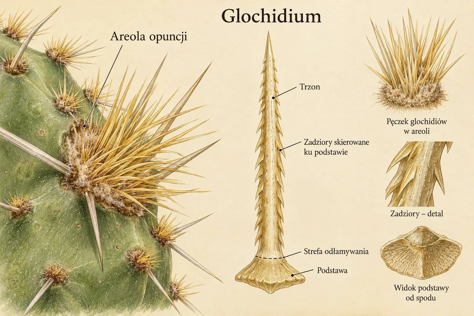
A mature spine contains dead fibres of strengthening tissue and a sclerotised epidermis. Fibres are elongated cells whose walls may occupy almost the entire cross-section or leave a broad lumen. A cactus spine is a modified leaf organ. Its mature tissue lacks stomata, photosynthetic parenchyma and its own vascular bundle. This structure defines how its function is analysed. [Mosco 2009, p. 120]
In 2009, Mosco compared spines of 30 species and lower-rank taxa historically assigned to Turbinicarpus. Plants were grown from documented seeds in the author's collection under dry conditions. Five spines were collected per taxon, one for electron microscopy, two for surface examination by light microscopy and two for sections. The resulting picture describes this collection material. [Mosco 2009, pp. 120–121]
Internal hardness and surface relief
In many hard spines, fibres with thick secondary walls and very narrow lumina occupied most of the interior. Thinner-walled cells could occur beneath the epidermis. Stiffness therefore depended on the organisation of the entire cross-section. A thin spine could have a bristle-like form even when it contained clearly thickened fibre walls. [Mosco 2009, pp. 121–123, 126]
Epidermal cells formed a smooth surface, short projections or longer outgrowths. Small conical projections were observed in Turbinicarpus pseudopectinatus. In T. laui, they were feathery. Kadenicarpus horripilus (Turbinicarpus horripilus in the paper) also had feathery outgrowths, although its central spine studied was hard and sharp. Epidermal relief and organ stiffness are therefore separate traits. [Mosco 2009, Mosco, pp. 121–122. Vázquez-Sánchez et al., p. 412] [Vázquez-Sánchez et al. 2019, Mosco, pp. 121–122. Vázquez-Sánchez et al., p. 412]
A papery ribbon and a corky structure
Papery spines have looser strengthening tissue, with relatively thin fibre walls and retained cell lumina. In Turbinicarpus schmiedickeanus subsp. flaviflorus (T. flaviflorus in the paper), the epidermis was continuous and smooth. Thin-walled fibres dominated the section studied. The subspecies rioverdensis (T. rioverdensis in the paper) had cork-textured spines with transverse fissures and a more varied fibre arrangement. [Mosco 2009, Mosco, p. 123, Table 2] [2-WWW]
In Turbinicarpus alonsoi and part of the T. schmiedickeanus complex, fissures passed through the epidermis and deeper tissue. Mosco related their formation to the brittleness of thin-walled fibres and changes during spine growth. Corky texture describes appearance. The spine tissue consists of fibres and epidermis, while cork on the older stem forms separately in the periderm. [Mosco 2009, pp. 123–127]
| Taxon studied | Spine description | Fibres in the deeper portion | Thin-walled fibres |
|---|---|---|---|
| T. valdezianus | Woody structure, feathery epidermis | 4,1 ± 0,9 | Not found |
| T. schmiedickeanus subsp. flaviflorus (T. flaviflorus in the paper) | Papery | Not found | 1,5 ± 0,3 |
| T. schmiedickeanus subsp. rioverdensis (T. rioverdensis in the paper) | Corky, with fissures | 3,1 ± 0,9 | 1,0 ± 0,2 |
Table values concern cells in preparations rather than the mean size of the entire spine. The author usually took 25 measurements of a trait, but when good sections were difficult to obtain, including in T. valdezianus, ten to twenty. The division of fibres in the source table describes their location and type in a given spine. Different tissue types can coexist in one organ. [Mosco 2009, p. 120, Table 2]
Feathery spines of the adult plant
Turbinicarpus valdezianus retains very long outgrowths of spine epidermal cells in adult specimens. Many other turbinicarpi studied have a feathery surface in the juvenile phase, after which the outgrowths of successive spines become shorter. A juvenile trait persisting in an adult plant is interpreted as neoteny. The comparison describes organ differentiation during development. [Mosco 2009, pp. 123, 126–127]
The dense feathery layer could shelter the stem surface from radiation. In the publication, this is a functional hypothesis linked to structure and habitat. The author did not measure the covering's light transmission, stem temperature or photosynthesis rate. In this species, microscopy showed thick internal fibre walls despite the spine's delicate external appearance. [Mosco 2009, pp. 122, 126–127]
Fissures and possible water movement
Breaks in the epidermis provide access to internal spine tissues. Mosco discusses earlier experiments in which a tracer applied to spines of Turbinicarpus schmiedickeanus subsp. klinkerianus was also detected in the stem near the areole. On this basis, he considers possible capillary water movement. This is a cited result from an older study combined with observation of spine structure. [Mosco 2009, p. 120]
Mosco's own study did not determine water flux or this pathway's contribution to plant supply. The question involves several stages, wetting the spine, movement of liquid through its tissues and entry into the living stem. Observing a fissure identifies a possible access route. The whole-plant balance requires quantitative assessment of successive stages. [Mosco 2009, pp. 120, 126–127]
The spine as a trait with several levels
The comparison distinguishes the shape of the entire spine, its epidermal relief and fibre-wall structure. A change at one level need not correspond to changes at the others. Bristle-like spines of T. laui and Kadenicarpus pseudomacrochele subsp. minimus (Turbinicarpus pseudomacrochele subsp. minimus in the paper) form with different wall thicknesses from the feathery spines of T. valdezianus. In these examples, a shared delicate appearance has different anatomical foundations. [Mosco 2009, Mosco, pp. 122–127. Vázquez-Sánchez et al., p. 412] [Vázquez-Sánchez et al. 2019, Mosco, pp. 122–127. Vázquez-Sánchez et al., p. 412]
The microscopic description can be related to qualitatively assessed properties of the material, its stiffness, texture and sharp or blunt tip. The study contains no tests of bending force, elasticity or tear resistance. Moving from a tissue image to a numerical description of mechanics requires further experimentation. The current result precisely shows how the same cell types form very different spines. [Mosco 2009, pp. 120–127]
Based on the full text of Mosco's 2009 paper. Table data were checked against page images. Current subspecies names follow POWO, with historical names in parentheses. Hypotheses about stem shelter and the contribution of spines to water relations retain the scope of the source evidence.
Micro-morphology and anatomy of Turbinicarpus (Cactaceae) spines
Mosco A., Micro-morphology and anatomy of Turbinicarpus (Cactaceae) spines, Revista Mexicana de Biodiversidad, 2009, 80(1):119-128, doi: 10.22201/ib.20078706e.2009.001.589
A study of spine surfaces and internal structure in 30 species and lower-ranked taxa from a documented collection. Light and electron microscopy show differences in fibre wall thickness, tissue arrangement and epidermal cell shape. Hard, bristle-like, papery and corky spines were compared, together with the persistent feathery hairs of T. valdezianus. Functions in stem protection and water uptake are discussed as hypotheses or results of earlier studies, without direct measurements in this experiment.
Polyphyly of the iconic cactus genus Turbinicarpus (Cactaceae) and its generic circumscription
Vázquez-Sánchez M., Sánchez D., Terrazas T., De la Rosa-Tilapa A., Arias S., Polyphyly of the iconic cactus genus Turbinicarpus (Cactaceae) and its generic circumscription, Botanical Journal of the Linnean Society, 2019, 190(4):405-420, doi: 10.1093/botlinnean/boz027
A phylogenetic analysis of the former broad circumscription of Turbinicarpus, combining sequences from five plastid DNA regions with morphological and anatomical traits. It recognises three distinct lineages, Turbinicarpus, Rapicactus and Kadenicarpus, and compares seed, fruit and stem tissue structure. It contains a reconstruction of range history, a generic key and a nomenclatural treatment of Kadenicarpus. Relationships among some species and subspecies remain poorly resolved.
Plants of the World Online — Royal Botanic Gardens, Kew
Accepted names, synonyms and distribution. A citation beside a taxon leads to its record.
Accessed 2026-10-08.