Botanical plates
New plates with panel explanations and references to the literature.
Schematic illustrations from the supplied materials. Captions identify the scope of simplifications and any inaccuracies. Clicking an image opens it at full size.
Showing 1–12 z 59 results

Stem cross-section
The plate shows the organisation of a fleshy stem. Green around the perimeter marks the photosynthetic region, and the pale tissue deeper inside marks the water storage region. The vascular system surrounds the pith. [Mauseth 2006]
This is a structural model. Tissue proportions, continuity of the hypodermal layer and distribution of additional bundles in the cortex vary among cacti.
Enlarge plate ↗
Areole and spine development
The left panel shows the positions of radial and central spines. The central panel connects the areole with stem tissues, and the right panel shows the elongated, hard cells of a mature spine. An areole is a short shoot that also produces hairs and other structures. [Mauseth 2006] [Silva et al. 2020]
The information about absence of xylem and phloem concerns ordinary spines. Specialised secretory glochids described in two species have a vascularised base and are shown on a separate plate.
Enlarge plate ↗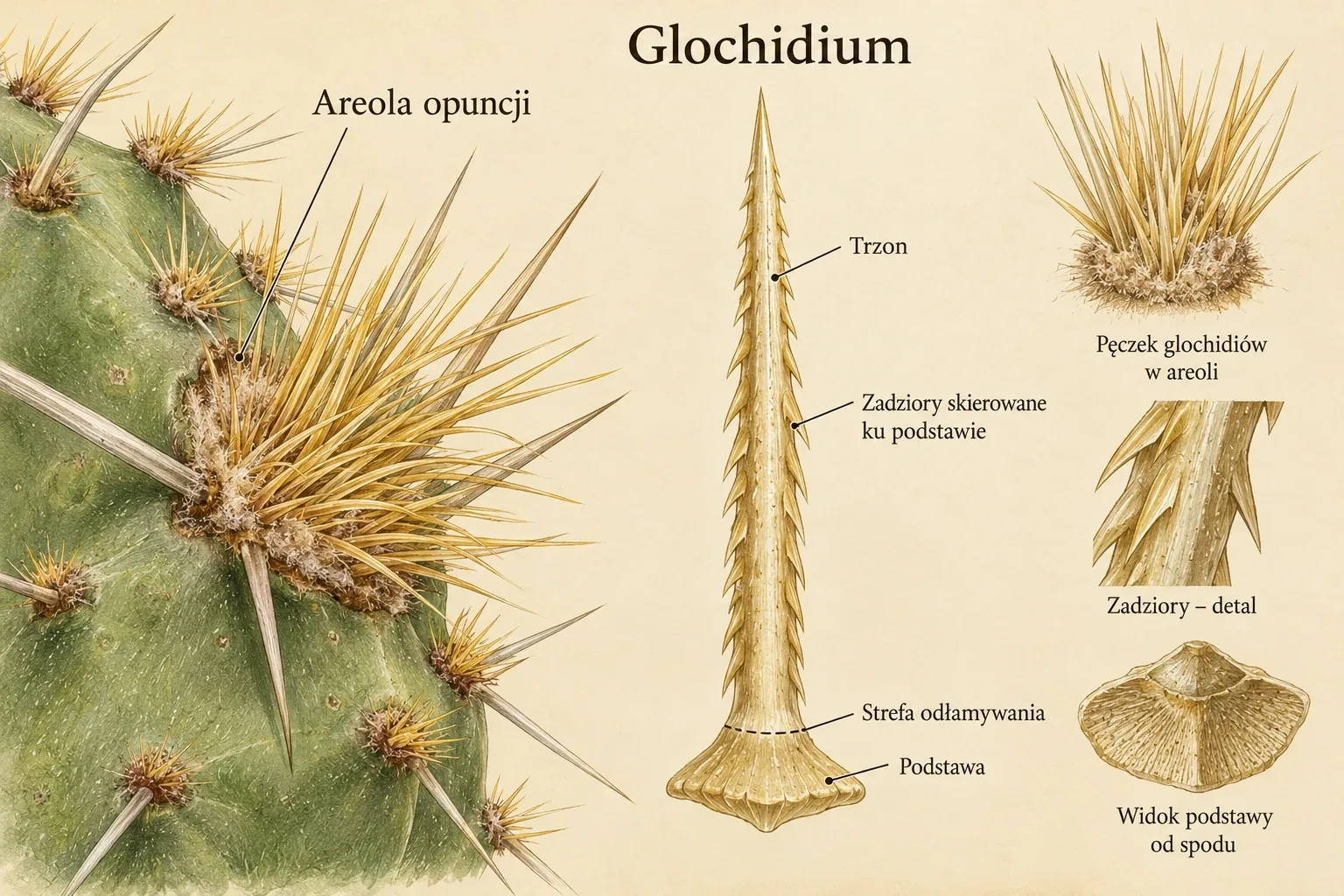
Opuntia glochidium
The large panel at left indicates the position of glochids in an areole. The central panel shows a single glochid with barbs pointing towards its base. At right are enlarged views of a bundle, the shaft surface and the base. [Robinson 1974]
The drawing is a schematic enlargement without a measurement scale. Base shape and the arrangement of surface cells are not identical in all opuntioids.
Enlarge plate ↗
Spine structure, distribution and types
The upper panels compare views of an areole, a single spine and its cross-section. Below are differences in spine shape and distribution within an areole. The terms “central” and “radial” describe position, while “hooked” and “flattened” describe shape. [Mauseth 2006] [Robinson 1974]
Categories may occur together on one plant. The plate is not a key for identifying species, and the cross-section proportions are schematic.
Enlarge plate ↗
Spines. Structure and possible functions
Panels 1–3 organise spine structure and descriptive traits. Panel 4 compares possible protective functions and effects on the environment around the stem surface. Panel 5 illustrates the developmental relationship between spines and leaves. [Mauseth 2006] [Robinson 1974] [Ju et al. 2012]
The arrow from leaf to spine indicates homology, not transformation of a mature leaf on a particular plant. The 100 μm scale bar is part of the drawing, with no measurement material specified. Spine effects on water and microclimate depend on species and conditions.
Enlarge plate ↗
Secretory glochids
The left side compares structures in Brasiliopuntia brasiliensis and Nopalea cochenillifera, accepted in the atlas as Opuntia cochenillifera. The right side distinguishes the expanded, vascularised base, unlignified middle part and tip. The drop at the tip indicates secreted nectar. [Silva et al. 2020]
The plate concerns specialised structures in two species studied. It does not show the structure of all glochids or ordinary spines. It is an educational reconstruction, not a microscopic image from a publication.
Enlarge plate ↗
Ribs, tubercles and stem form
Two rows of panels compare rib direction, number, width and degree of division into tubercles. These traits help describe stem appearance and changes during growth. [Mauseth 2006] [Hernández-Hernández et al. 2011]
The plate labels combine different descriptive criteria. Segmentation of the whole stem is not a rib type, and tubercles do not form a single simple evolutionary series from ribs. This comparison is not a formal classification.
Enlarge plate ↗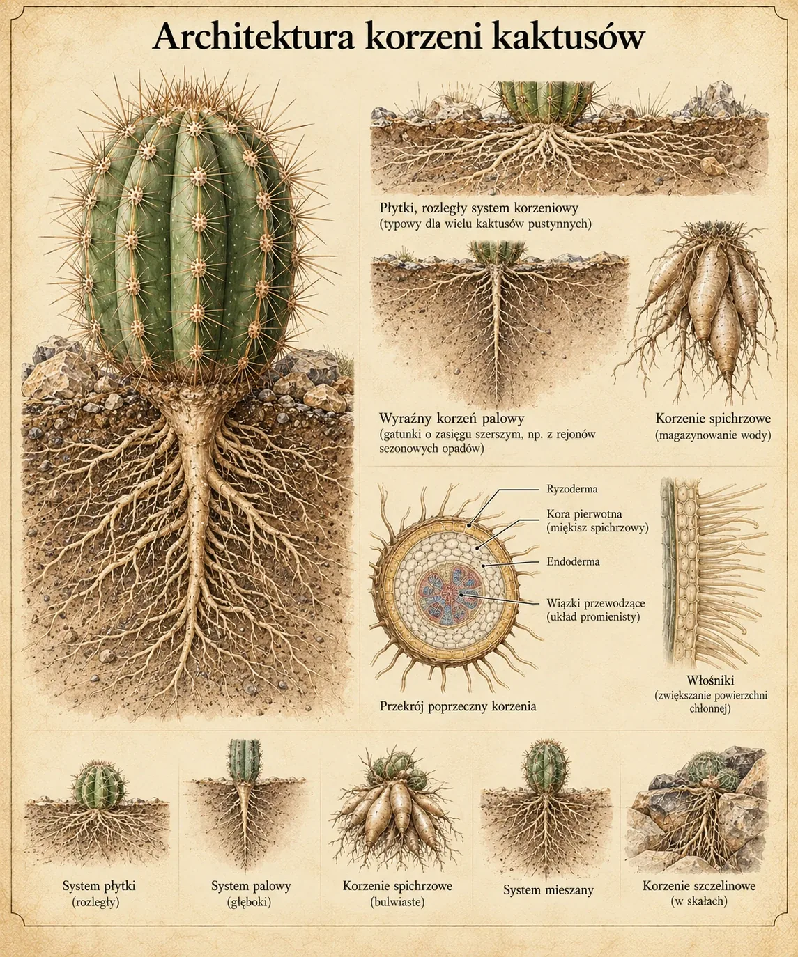
Root architecture
The plate combines a whole-plant view with a comparison of root spread and thickening. Two smaller panels show the general route from the root surface through the cortex and endodermis to vascular tissues, and the role of root hairs in contact with the substrate. [Dubrovsky and North 2002] [Dubrovsky et al. 1998]
Internal structure is shown schematically. A taproot is not always deep, and “crevice” describes its position in the substrate. Architecture also depends on age, rainfall, damage and container conditions.
Enlarge plate ↗
Six views of root systems
The upper row compares an extensive shallow system, a dominant main root and roots that store resources. The lower row shows a mixed system, growth in crevices and shallow spreading Opuntia roots. [Dubrovsky and North 2002]
These are examples of architecture, not six mutually exclusive taxonomic categories. A plant may combine several of the traits shown, and lengths and depths are not drawn to scale.
Enlarge plate ↗
Flower, ovary, fruit and seeds
The central drawing identifies the perianth, stamens, stigma, style and ovary. Side panels cover the flower interior, pollen, fruit and seeds. The lower strip compares general flower shapes. [Rosas-Reinhold et al. 2021] [Ramírez-Castro et al. 2024] [González et al. 2023]
The plate requires anatomical correction when interpreting details. The partitions drawn in the ovary section do not represent the unilocular ovary with parietal placentation typical of cacti. Stamens surround the style and are not part of it. The pollen and seed scales and the fruit shown do not document a single identified species.
Enlarge plate ↗
Structures developing in an areole
An areole is a strongly shortened lateral shoot. The plate compares a young areole, spine formation, flower bud development and an areole with mature spines. The panels help distinguish structures but do not define a mandatory order of appearance. In some cacti, a period of meristem dormancy separates spine formation from flowering. [Mauseth 2006]
The label “meristem” refers to the formative tissue, not the whole visible areole cushion. The 5 mm scale bar is not linked to an identified species or a measurement. The figure does not document an experiment or development time.
Enlarge plate ↗
Areoles, buds and a flower on the stem
The drawing shows areole distribution on the ribs and their connection with spines and lateral stem structures. An areole meristem may participate in the development of a branch or floral structure. A closed bud is shown next to an open flower. These are different structures shown in one diagram, without assignment to an identified species. [Mauseth 2006] [Rosas-Reinhold et al. 2021]
The label “spines” refers to the elongated, sharply pointed elements, not the pale hairs in the centre of the areole. The green bud labelled “offset” represents the drawing's intended interpretation. Its outline alone does not document developmental identity or internal structure.
Enlarge plate ↗