Nectar on a young stem segment
In Opuntia robusta, nectar appears outside flowers in the areoles of young segments. In 2018, Sandoval-Molina and colleagues described the producing structures using light, scanning and transmission electron microscopy. Their position and successive developmental stages indicate that they are modified spines. [Sandoval-Molina et al. 2018]
In the young areoles studied, glands lay among other armature elements, behind the leaf and main spine. They were small, conical and active for a limited growth period. This position links secretion with the youngest stem region before its tissues finish maturing. [Sandoval-Molina et al. 2018]
Three zones of one organ
At the base was a meristematic zone whose dividing cells build the organ. Above it lay an elongation zone, and secretory tissue formed the apex. Surface cells in this zone were convex and covered by a thin, folded cuticle. Anatomy thus allows the transition from growth to secretory specialisation to be followed. [Sandoval-Molina et al. 2018]
No vascular bundles were found inside the nectary. Vascular tissues reached beneath its base, and substances passed onwards through living cells. Plasmodesmata, cytoplasmic connections, were visible in the walls between them. Determining the exact contributions of transport pathways requires physiological measurements. Sections reveal their anatomical basis. [Sandoval-Molina et al. 2018]
A cell prepared for secretion
Cells of the apical zone contained numerous mitochondria, Golgi apparatuses, endoplasmic reticulum components and vesicles. This set of organelles corresponds to intense metabolism and the production and movement of substances. Transmission microscopy also showed differences between cells at successive nectary developmental stages. [Sandoval-Molina et al. 2018]
The authors considered secretion release through small breaks or openings in the cuticle and a contribution from ants damaging the surface. As it matured, the spine lignified and secretory activity ceased. The same organ thus passed through a metabolically active phase, followed by an increased contribution of lignin-stiffened walls. [Sandoval-Molina et al. 2018]
Ants at the food source
Ants of the genera Camponotus and Forelius visited young segments. In a one-time plant comparison, more ants were found on younger, narrower segments. The relationship corresponded to the distribution of active nectaries. The authors also observed secretion consumption and aggressive behaviour towards objects touching the stem. [Sandoval-Molina et al. 2018]
The experiment did not include ant exclusion or long-term measurement of herbivore damage. The gland, its development and the insects using it were therefore documented. A protective effect for the prickly pear remains to be tested by comparing plants with and without ant access, taking actual herbivore pressure into account. [Sandoval-Molina et al. 2018]
The areole as a group of specialised organs
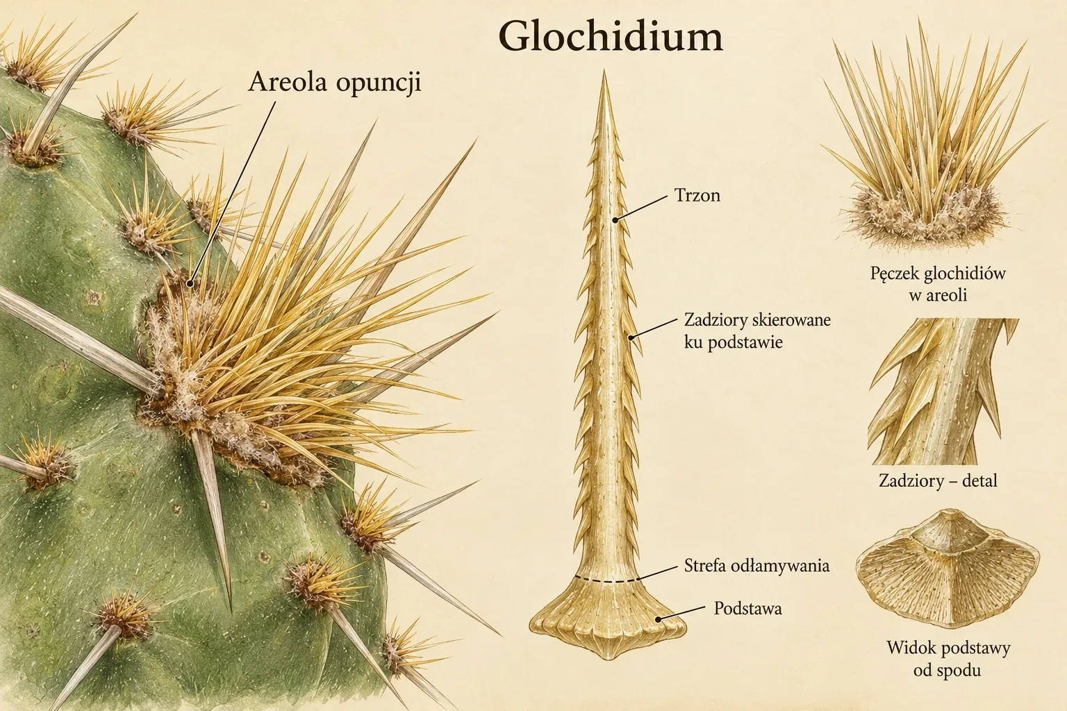
The case of O. robusta shows how spines, glochids, hairs and a short-lived secretory structure coexist at one stem site. Living cells, a thin surface and developmental stage determine its function. The organ's spine identity and ability to secrete nectar belong to two complementary descriptive levels, origin and activity. [Sandoval-Molina et al. 2018]
Prepared from the full texts of the cited publications. Taxon names follow the current POWO treatment, with names used in the studies retained.
First description of extrafloral nectaries in Opuntia robusta (Cactaceae): Anatomy and ultrastructure
Sandoval-Molina M. A., Zavaleta-Mancera H. A., León-Solano H. J., Solache-Ramos L. T., Jenner B., Morales-Rodríguez S., Patrón-Soberano A., Janczur M. K., First description of extrafloral nectaries in Opuntia robusta (Cactaceae): Anatomy and ultrastructure, PLoS ONE, 2018, 13(7):e0200422, doi: 10.1371/journal.pone.0200422
The first detailed description of extrafloral nectaries in Opuntia robusta, including their development as modified spines, anatomy and secretory cell ultrastructure. It is complemented by observations of ants on young cladodes and changes in nectaries during maturation.